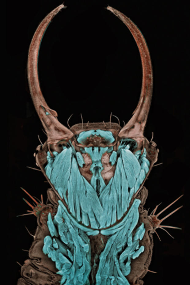
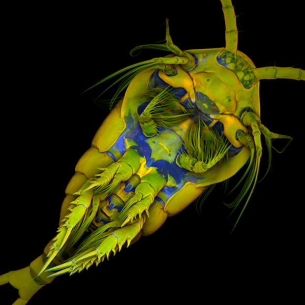
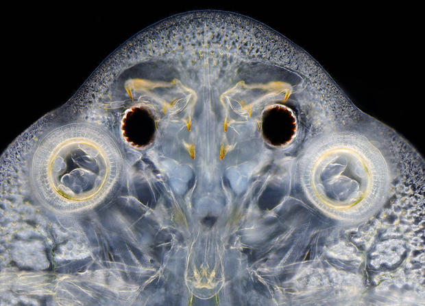
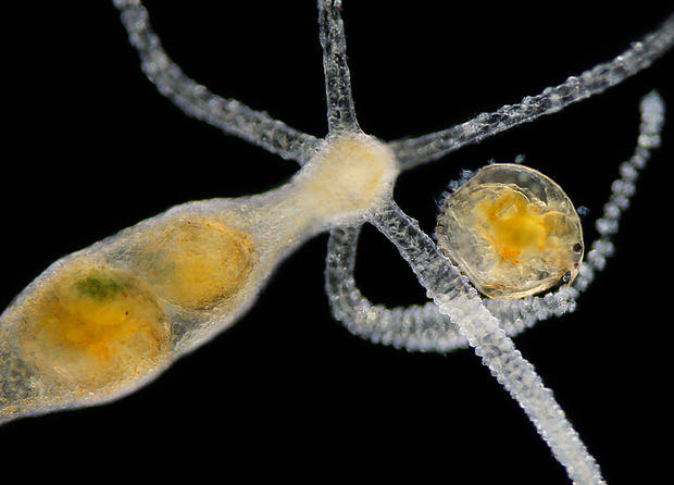
Dr. Igor Siwanowicz, Max Planck Institute of Neurobiology
The Nikon International Small World Competition showcases micro-photographic images that are not simply astounding capture of nature or science on a microscopic level; they are also beautifully composed and colorful depictions of the very structures of life itself.
The competition, which began in 1974, celebrates the work of photomicrographers, amateur and professional alike, from a wide array of scientific disciplines. Nikon recently announced its 2011 award-winners.
At left is this year's first place winner: Portrait of a Chrysopa sp. (green lacewing) larva (20x) by Dr. Igor Siwanowicz, of the Max Planck Institute of Neurobiology in Martinsried, Germany.
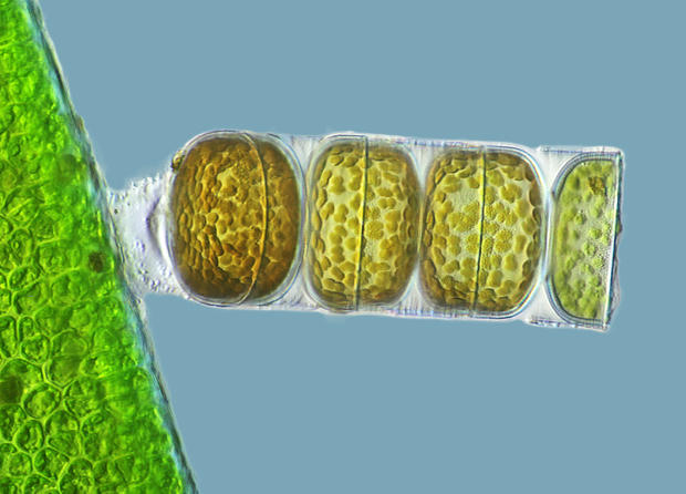
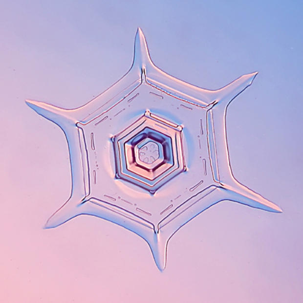
Frank Fox, Fachhochschule Trier
3rd Place: A living specimen of Melosira moniliformis (320x) by Frank Fox, Fachhochschule Trier, Trier, Rheinland-Pfalz, Germany.
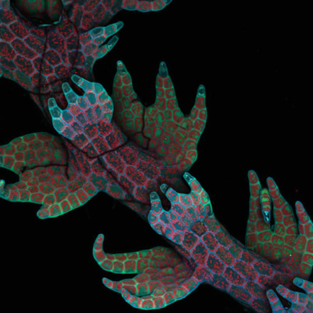
Dr. Robin Young, University of British Columbia
4th Place: Dr. Robin Young of the University of British Columbia in Vancouver captured the intrinsic fluorescence in Lepidozia reptans (liverwort) via confocal microscopy (20x).
Dennis Callahan, CIT
6th Place: Dennis Callahan of the California Institute of Technology in Pasadena photographed cracked gallium arsenide solar cell films (50x).
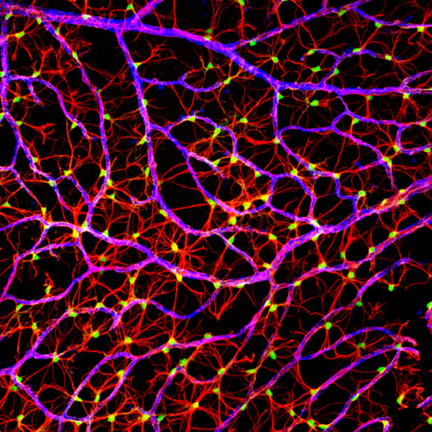
Gabriel Luna, UC Santa Barbara
7th Place: This laser confocal scan of the retinal flatmount of a mouse nerve fiber layer (40x) was photographed by Gabriel Luna of UC Santa Barbara's Neuroscience Research Institute.
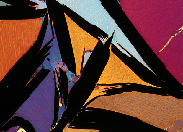
Dr. Bernardo Cesare
8th Place: This colorful image, shot with polarized light, captures graphite-bearing granulite from Kerala, India (2.5x), was created by Dr. Bernardo Cesare of the Department of Geosciences in Padova, Italy.
Dr. Jan Michels
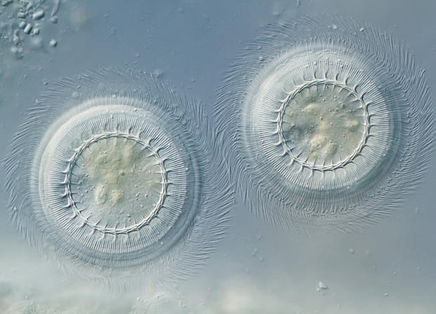
9th Place: A ventral view of Temora longicornis (marine copepod) (10X), photographed via confocal, autofluorescence and congo red fluorescence by Dr. Jan Michels, at Christian-Albrechts-Universit
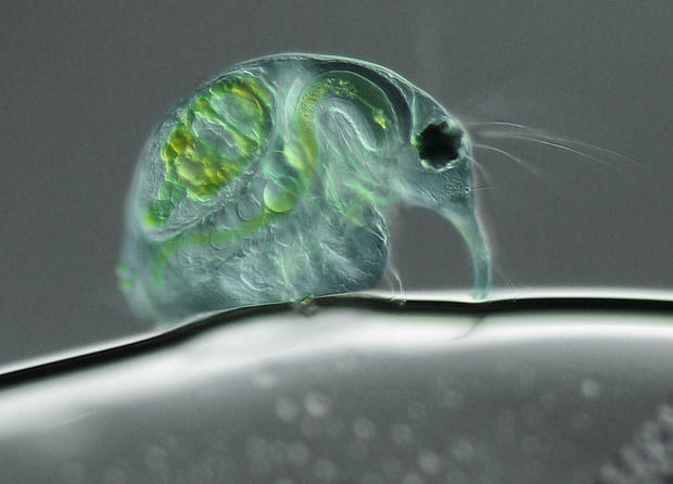
Joan Rohl
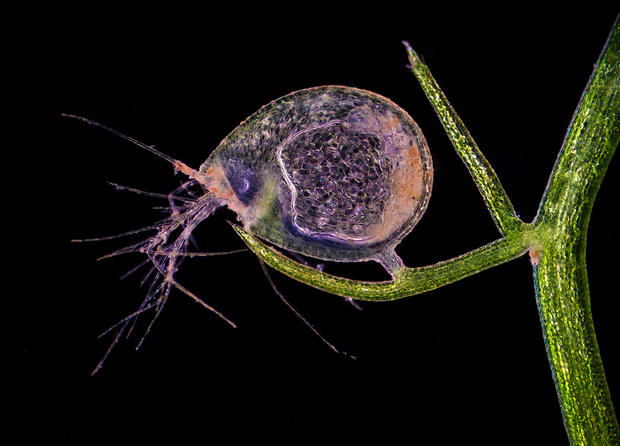
10th Place: A 100-magnification picture of a freshwater water flea (Daphnia magna) was taken by Joan Rohl of the Institute for Biochemistry and Biology in Potsdam, Germany.
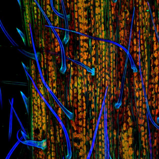
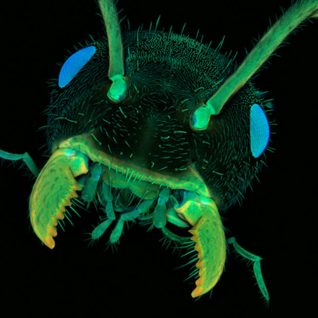
Dr. Jan Michels
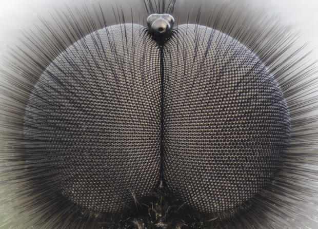
11th Place: A confocal autofluorescence image of an ant head (10x), by Dr. Jan Michels of the Christian-Albrechts-Universit
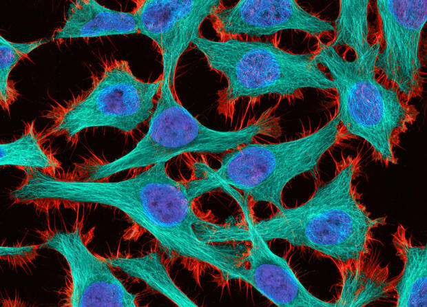
Thomas Deerinck, National Center for Microscopy and Imaging Research
12th Place: HeLa (cancer) cells, at 300 magnification, as photographed via 2-Photon fluorescence by Thomas Deerinck at the National Center for Microscopy and Imaging Research in La Jolla, Calif.
Dr. Stephen S. Nagy, Montana Diatoms
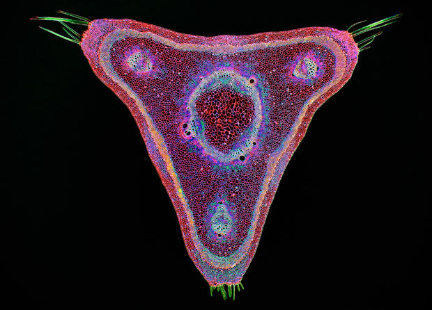
13th Place: A brightfield, digitally inverted cross-section of a curare vine (Chondrodendron tomentosum) at 45x magnification, photographed by Dr. Stephen S. Nagy of Montana Diatoms in Helena, Mont.
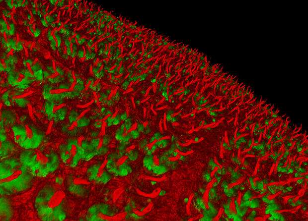
James H. Nicholson/NOAA/NOS/NCCOS/CCEHBR & HML
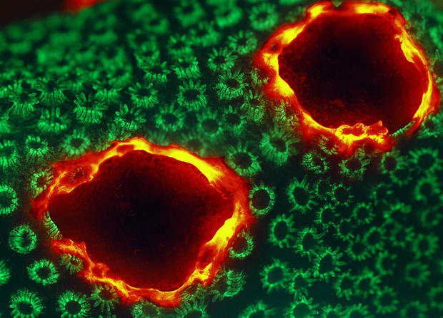
15th Place: A live specimen of Porites lobata (lobe coral), displaying tissue pigmentation response with red fluorescence (12x). Photograph by James H. Nicholson of the Coral Culture and Collaborative Research Facility in Charleston, S.C.
Dr. Christopher Guerin
16th Place: Dr. Christopher Guerin of the Flanders Institute of Biotechnology in Ghent, Belgium, photographed cultured cells growing on a bio-polymer scaffold (63x).
Dr. Witold Kilarski
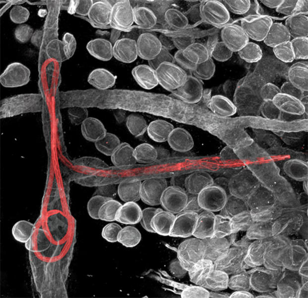
17th Place: A fluorescent confocal microscopic photo of Litomosoides sigmodontis (filaria worms) inside lymphatic vessels of the mouse ear (150x), by Dr. Witold Kilarski of the EPFL-Laboratory of Lymphatic and Cancer Bioengineering in Lausanne, Switzerland.
Benjamin Blonder and David Elliott
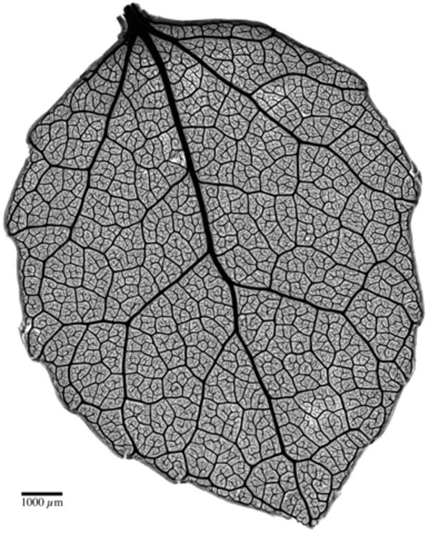
18th Place: A brightfield image of safranin-stained tissue revealing the venation network of a young Populus tremuloides (quaking aspen) leaf (4x) by Benjamin Blonder and David Elliott of the University of Arizona in Tucson.
Dr. Donna Stolz, University of Pittsburgh
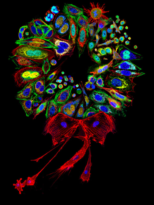
19th Place: A mammalian cell collage stained for various proteins and organelles, assembled into a wreath (200-2,000x) by Dr. Donna Stolz, University of Pittsburgh.
Douglas Moore, UW-Stevens Point
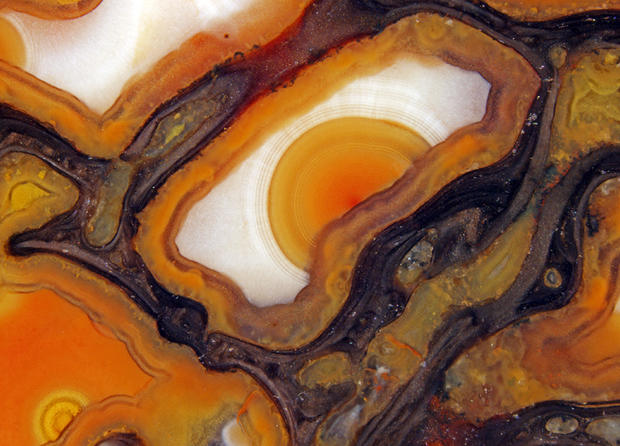
20th Place: Douglas Moore of the University of Wisconsin-Stevens Point used stereomicroscopy, and fiber optics to capture agatized dinosaur bone cells, unpolished, ca. 150 million years old (42x).
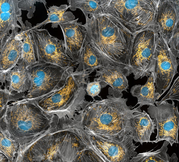
Dr. Torsten Wittmann, UCSF
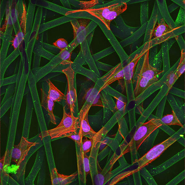
A 60x-mag image of bovine pulmonary artery endothelial (BPAE) cells, fixed and stained for actin, mitochondria and DNA, incorporating epi-fluorescence and multi-image stitching, by Dr. Torsten Wittmann of the University of California, San Fransisco.
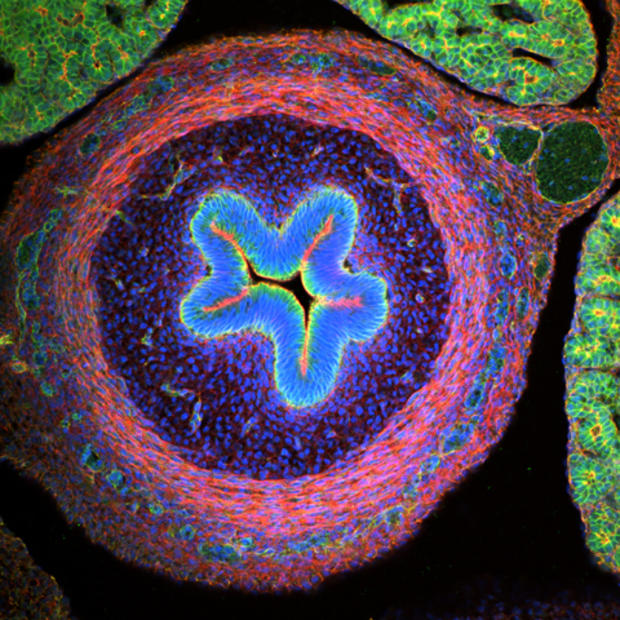
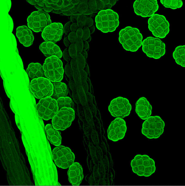
Poulomi Ray, Clemson University
Poulomi Ray of the Department of Biological Sciences at Clemson University in Clemson, S.C., photographed a chick embryo intestine (20X).
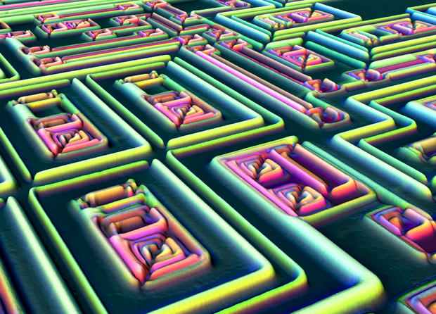
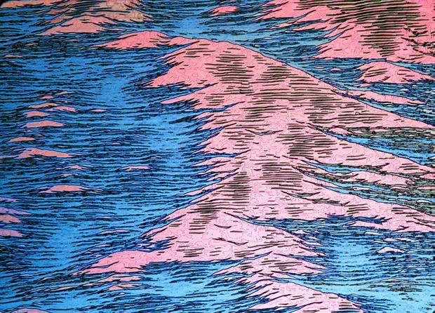
Dr. Pedro Barrios-Perez/NRCC/IMS
This landscape is actually the surface of a gallium antimonide semiconductor wafer after metal peel-off (200x), photographed by Dr. Pedro Barrios-Perez, National Research Council of Canada/Institute for Microstructural Sciences, Ottawa, Ontario.
Dr. Paul Appleton, University of Dundee
Stem cells of a mouse's small intestine (40x), by Dr. Paul Appleton of the University of Dundee in Dundee, Scotland.
Dr. Marta Guervos, University of Oviedo
Autofluorescence colors this 40x image of Acacia dealbata (Silver Wattle) pollen grains, by Dr. Marta Guervos, Image Processing Unit, Scientific-Technical Facilities, University of Oviedo in Asturias, Spain.
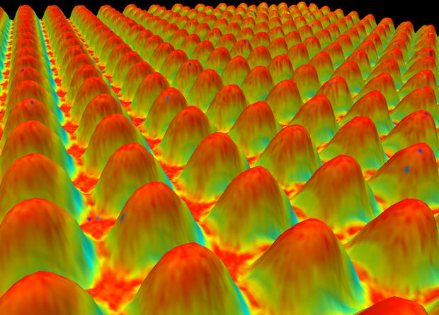
Kevin Smith, MetPrep Ltd.
Kevin Smith of MetPrep Ltd. in Warwickshire, U.K., photographed a charge coupled device (CCD) sensor used in digital imaging (1000x).
Jose R. Almodovar
Jose R. Almodovar of the Microscopy Center, Biology Department, UPR Mayaguez Campus in Mayaguez, Puerto Rico, photographed the Utricularia gibba (bladderwort) (40x).
Dr. Jorge Bernardino de la Serna
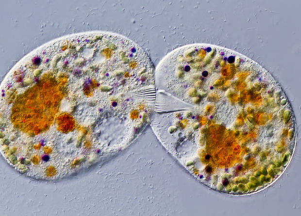
Dr. Jorge Bernardino de la Serna of MEMPHYS - Center for Biomembrane Physics, Department of Biochemistry and Molecular Biology in Odense, Denmark, produced this 40x image of giant liposomes of pulmonary surfactant.
For more on Nikon's 2011 Small World Competition and to view other entries visit the Nikon Small World Competition website.