Amazing tiny things
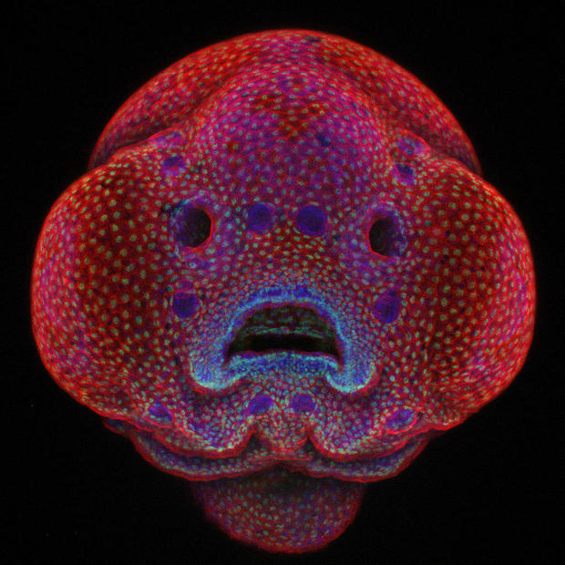
A microscopic image of a fish face by Oscar Ruiz took first prize in the 42nd Annual Nikon Small World Photomicrography Competition in 2016. The contest comprised fascinating submissions from 70 countries submitted by scientists, photographers as well as hobbyists.
Ruiz, who works at The University of Texas MD Anderson Cancer Center in Houston, took the winning image as part of his research on the facial development of a four-day-old zebrafish embryo. His research, with time-lapse photography playing an integral role, is focused on studying gene mutations that lead to facial abnormalities such as cleft lip and palate in humans. His image proves that images can be both useful to science and beautiful to look at too.
“Using a live-imaging approach means we can better understand and pinpoint exactly how and why these developmental abnormalities occur. The first step is knowing how it happens, then we can figure out how to fix it,” explains Ruiz.
Take a look at the top images from the contest that showcase a microscopic world.
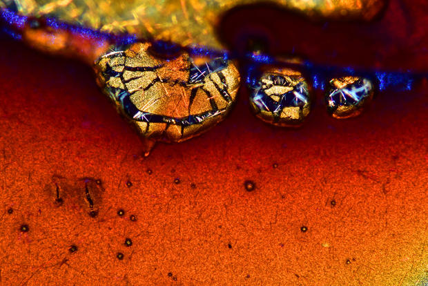
2nd Place
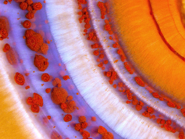
A polished slab of Teepee Canyon agate
Douglas L. Moore, University of Wisconsin Museum of Natural History
3rd Place
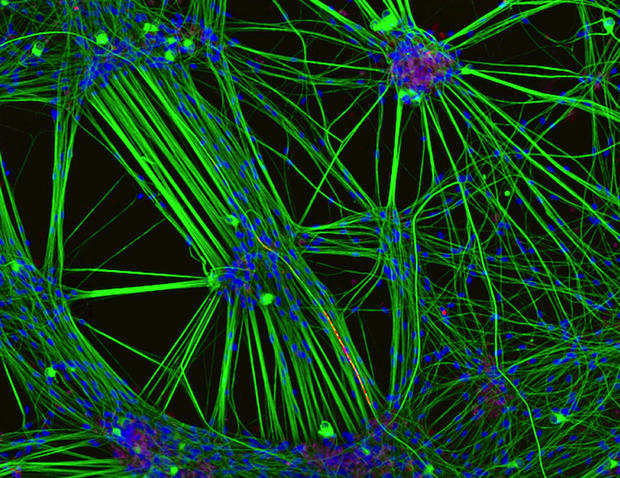
A culture of neurons (stained green) derived from human skin cells, and Schwann cells, a second type of brain cell (stained red)
Rebecca Nutbrown, University of Oxford, United Kingdom
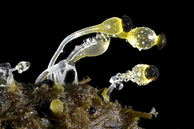
4th Place
Butterfly proboscis
Jochen Schroeder, Chiang Mai, Thailand
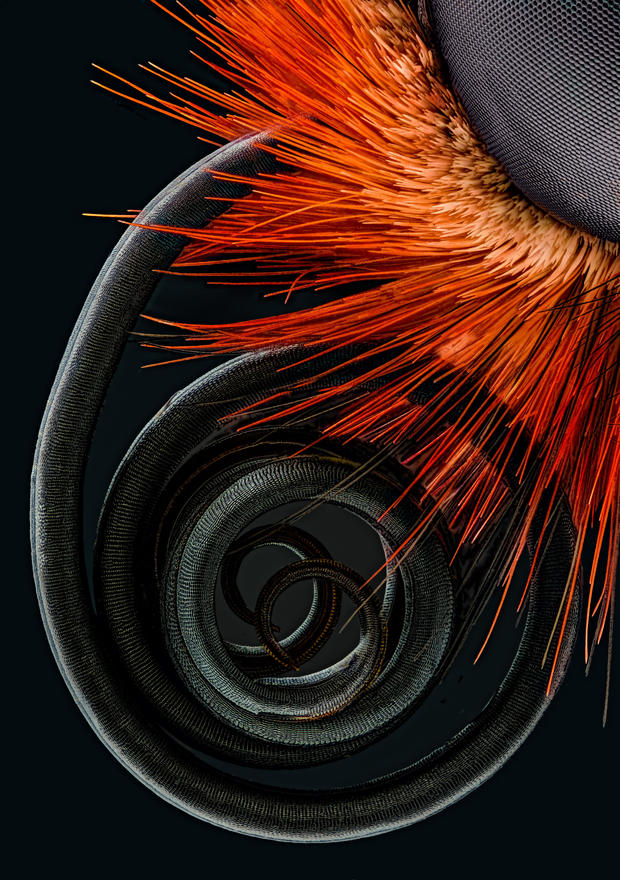
5th Place
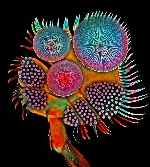
Front foot (tarsus) of a male diving beetle
Dr. Igor Siwanowicz, Howard Hughes Medical Institute (HHMI), Ashburn, Virginia, USA
6th Place
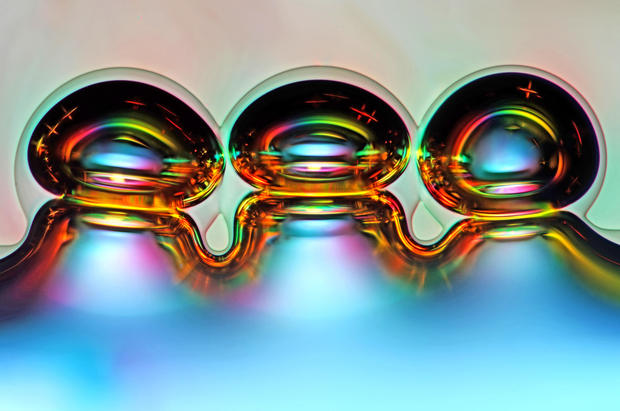
Air bubbles formed from melted ascorbic acid crystals
Marek Mis, Suwalki, Podlaskie, Poland
7th Place
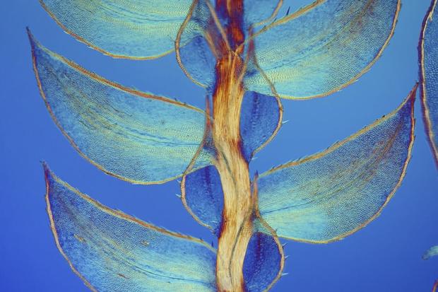
Leaves of Selaginella (lesser club moss)
Dr. David Maitland, Feltwell, United Kingdom
8th Place
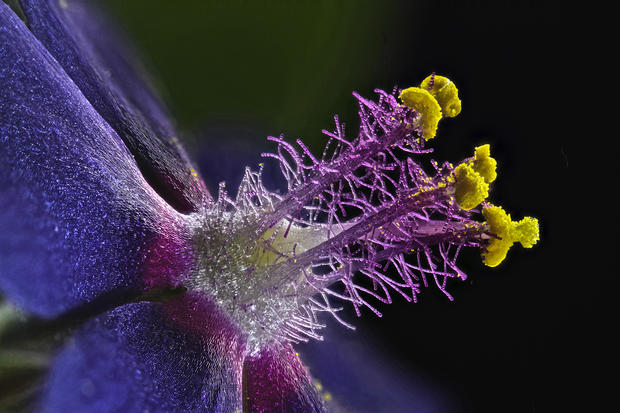
Wildflower stamens
Samuel Silberman, Monoson Yahud, Israel
9th Place
Espresso coffee crystals
Vin Kitayama and Sanae Kitayama, Azumino, Nagano, Japan
10th Place
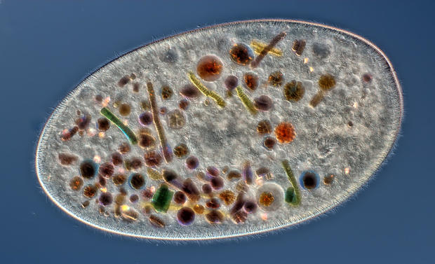
Frontonia (showing ingested food, cilia, mouth and trichocysts)
Rogelio Moreno Gill, Panama, Panama
11th Place
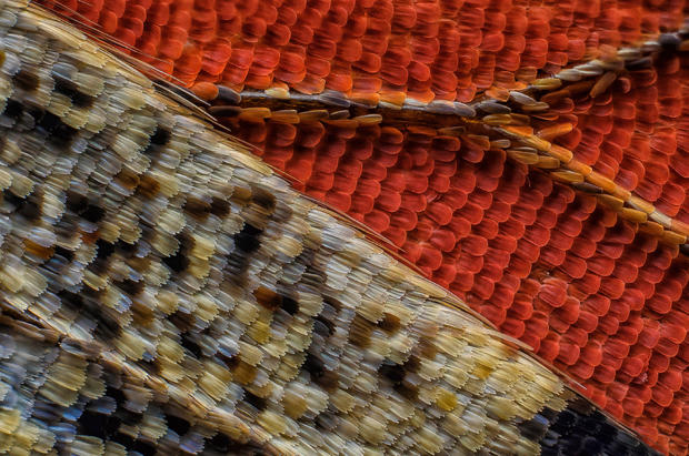
Scales of a butterfly wing underside (Vanessa atalanta)
Francis Sneyers, Brecht, Belgium
12th Place
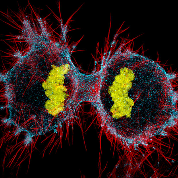
Human HeLa cell undergoing cell division (cytokinesis)--DNA (yellow), myosin II (blue) and actin filaments (red)
Dr. Dylan Burnette, Vanderbilt University School of Medicine, Nashville,
13th Place
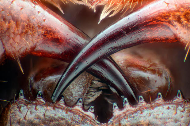
Poison fangs of a centipede (Lithobius erythrocephalus)
Walter Piorkowski, South Beloit, Illinois
14th Place
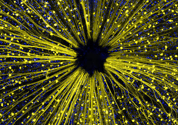
Mouse retinal ganglion cells
Dr. Keunyoung Kim, University of California, San Diego
15th Place
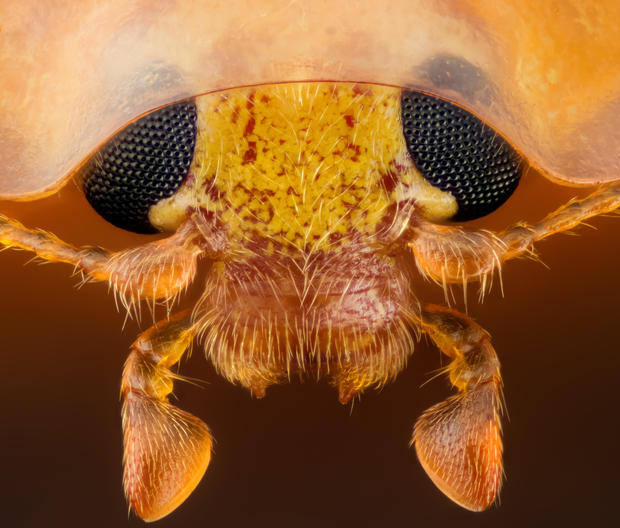
Head section of an orange ladybird (Halyzia sedecimguttata)
Geir Drange, Asker, Norway
16th Place
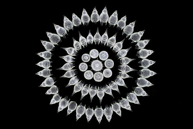
Sixty five fossil Radiolarians (zooplankton) carefully arranged by hand in Victorian style
Stefano Barone, Palazzo Pignano, Italy
17 Place
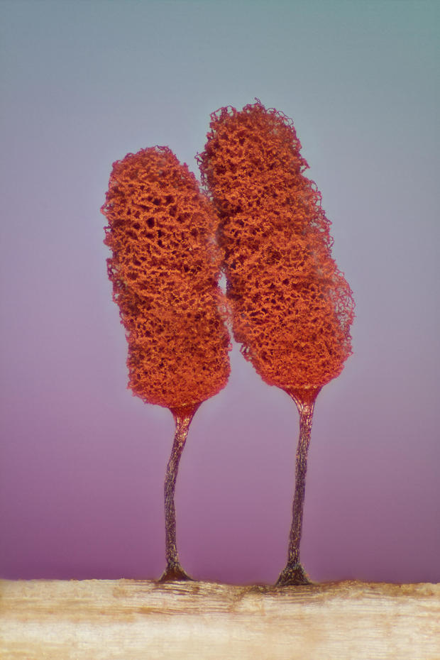
Slime mold (Mixomicete)
Jose Almodovar, University of Puerto Rico, Biology Department
18th Place
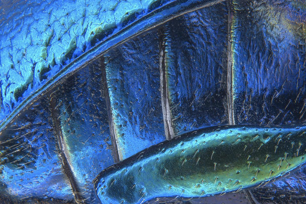
Parts of wing-cover (elytron), abdominal segments and hind leg of a broad-shouldered leaf beetle (Oreina cacaliae)
Pia Scanlon, Department of Agriculture and Food, Western Australia, Biosecurity and Regulation, South Perth
19th Place
Human neural rosette primordial brain cells, differentiated from embryonic stem cells
Dr. Gist F. Croft, Lauren Pietilla, Stephanie Tse, Dr. Szilvia Galgoczi, Maria Fenner, Dr. Ali H. Brivanlou, Rockefeller University, New York City
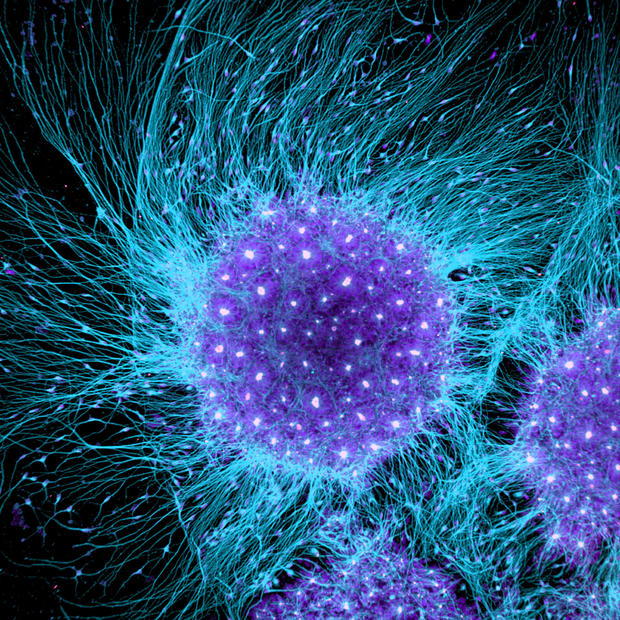
20th Place
Cow dung
Michael Crutchley, Haverfordwest, Pembrokeshire, United Kingdom
For more great photos: On Instagram - @nikoninstruments