Book excerpt: "The Song of the Cell" by Siddhartha Mukherjee
Dr. Siddhartha Mukherjee, the Pulitzer Prize-winning author of "The Emperor of All Maladies: A Biography of Cancer," returns with "The Song of the Cell: An Exploration of Medicine and the New Human" (Scribner), the history of the discovery of cells, and the science of manipulating them.
Read an excerpt below, and don't miss Kelefa Sanneh's interview with Siddhartha Mukherjee on "CBS Sunday Morning" October 30!
The Visible Cell
"Fictitious Stories About the Little Animals"
In the sum of the parts, there are only the parts.
The world must be measured by eye.
—Wallace Stevens
"The world must be measured by eye."
Modern genetics was launched by the practice of agriculture: the Moravian monk Gregor Mendel discovered genes by cross-pollinating peas with a paintbrush in his monastery garden in Brno. The Russian geneticist Nikolai Vavilov was inspired by crop selection. Even the English naturalist Charles Darwin had noted the extreme changes in animal forms created by selective breeding. Cell biology, too, was instigated by an unassuming, practical technology. Highbrow science was born from lowbrow tinkering.
In the case of cell biology, it was simply the art of seeing: the world measured, observed, and dissected by the eye. In the early seventeenth century, a Dutch father and son team of opticians, Hans and Zacharias Janssen, placed two magnifying lenses on the top and bottom of a tube and found that they could magnify an unseen world.* Microscopes with two lenses would be eventually termed "compound microscopes," while those with single lenses were called "simple"; both relied on centuries of innovation in glassblowing that had made its way from the Arabic and Greek worlds to the workshops of Italian and Dutch glassmakers. In the second century BC, the writer Aristophanes described "burning globes": spheres of glass sold as baubles in the market to concentrate and direct beams of light; if you looked carefully through a burning globe, you might see that same miniature universe magnified. Stretch that burning globe into an eye-sized lens, and you get the spectacle—invented supposedly by an Italian glassmaker, Amati, in the twelfth century. Mount it on a handle, and you have a magnifying glass.
* Some historians have argued that the Janssens' competitors, eyeglass makers Hans Lipperhey and Cornelis Drebbel, invented the compound microscope independently. The dates of all these inventions are in dispute, but likely occurred sometime between the 1590s and the 1620s.
The crucial innovation introduced by the Janssens was to fuse the art of glassblowing to the engineering of moving the pieces of glass on a mounted plate. By assembling one or two perfectly lucid pieces of lens-shaped glass on metal plates or tubes, with systems of screws and cogs to slide them, scientists would soon find their way into an unseen, miniature world—a whole cosmos previously unknown to humans—the obverse of the macroscopic cosmos observable through a telescope.
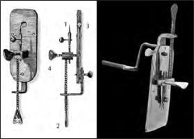
A secretive Dutch trader had taught himself to visualize this invisible world. In the 1670s, Antonie van Leeuwenhoek, a cloth merchant in Delft, needed an instrument to examine the quality and integrity of thread. Seventeenth-century Netherlands was a booming nexus of cloth merchandising—silks, velvets, wool, linen, and cotton came in swaths and bundles from ports and colonies, and were traded via the Netherlands throughout continental Europe. Building on the Janssens' work, Leeuwenhoek built himself a simple microscope, with a single lens secured on a brass plate, and a tiny stage to mount the specimens. At first, he used it to grade the quality of cloth. But his interest in his handmade instrument soon turned compulsive: he focused his lens on whatever objects he could find.
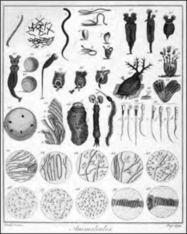
On May 26, 1675, the city of Delft was inundated by a storm. Leeuwenhoek, then forty-two, gathered some of the water from the drains of his rooftop, let it stand for a day, and then put a droplet under one of his microscopes and held it up to the light. He was instantly entranced. No one he knew had seen anything like it. The water was roiling with dozens of kinds of tiny organisms—"animalcules," he called them. Telescopists had seen macroscopic worlds—the blue-tinged moon, gaseous Venus, ringed Saturn, red-flecked Mars—but no one had reported a marvelous cosmos of a living world in a raindrop. "This was to me among all the marvels that I have discovered in nature the most marvelous among them all," he wrote in 1676. "No greater pleasure has yet come to my eye than these spectacle of the thousands of living creatures in a drop of water."*
* Leeuwenhoek had observed the presence of microscopic, single-celled organisms as early as 1674, but his letter to the Royal Society, dated 1676, had the most vivid descriptions of such organisms in standing rainwater.
He wanted to look more, to build finer instruments to visualize this captivating new universe of living beings. And so Leeuwenhoek purchased the highest-quality beads and globules of Venetian glass and then ground and polished them laboriously into perfectly lenticular shapes (some of his lenses, we now know, were made by stretching a rod of glass into a thin needle on a live flame, breaking the end, and then letting the needle "bubble" into a lens-shaped globule). He mounted these lenses on thin metal plates, crafted of brass, silver, or gold, each with an increasingly complex system of miniature armatures and screws to move parts of the instrument up and down and attain perfect focus. He made nearly five hundred such scopes, each a marvel of meticulous tinkering.
Were such creatures present in other samples of water as well? Leeuwenhoek entreated a man who was traveling to the seaside to bring him back a sample of ocean water in a "clean glass bottle." And again he found tiny single-celled organisms—"the body of a Mouse Color, clear towards the oval point"—swimming in the water. Eventually, in 1676, he recorded his findings and sent them to the most august scientific society of its time.
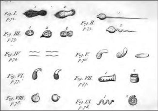
"In the year 1675," he wrote to the Royal Society of London, "I discover'd living creatures in Rain water, which had stood but few days in a new earthen pot. When these animalcula or living atoms did move, they put forth two horns, continually moving themselves. The rest of the body was roundish, sharpening a little toward the end, where they had a tail, nearly four times the length of the body."
By the time I'd finished writing that last paragraph, I was similarly obsessed: I wanted to look as well. Suspended in mid-pandemic limbo, I chose to build my own microscope, or at least the closest version that I could create. I ordered a metal plate and a turning knob, drilled a hole, and mounted the plate with the best tiny lens I could buy. It looked as much like a modern microscope as a bullock cart resembles, say, a spaceship. I trashed dozens of prototypes until I finally had one that might work. On a sunny afternoon, I placed a droplet of stagnant rainwater from a puddle on the mounting pin and held the apparatus up to the sunlight.
Nothing. Hazy forms, like shadows from a ghostly world, moved across my field of vision. A blur. Disappointed, I adjusted the focusing knob gently, as Leeuwenhoek would have. The anticipation made me feel each turn of the screw viscerally, as if the knob were, in fact, twisting its way up my spine. And suddenly I could see. The drop came sharply into view, and then a whole world within it. An amoeboid form flashed across the lens. There were branches of an organism I could not name. Then a spiral organism. A round, moving blob, surrounded by a halo of the most beautiful, the most tender filaments that I had ever seen. I could not stop seeing. Cells.
In 1677, Leeuwenhoek observed human spermatozoa, "a genital animalcule," in his semen as well as in a sample from a man with gonorrhea. He found them "moving like a snake or an eel swimming in water." Yet despite his ardor and productivity, the cloth merchant was notoriously reluctant to let observers or scientists examine his instruments. The suspicion was reciprocal, as scientists were often just as dismissive of him. Henry Oldenburg, the secretary of the Royal Society, implored Leeuwenhoek to "acquaint us with his method of observing, that others may confirm such Observations as these," and to provide drawings and confirmatory data, for of the roughly two hundred letters that Leeuwenhoek sent to the society, only about half offered evidence or used scientific methods considered fit for publication. But Leeuwenhoek would provide only vague details of his instruments or his methods. As the science historian Steven Shapin wrote, Leeuwenhoek was "neither a philosopher, a medical man, nor a gentleman. He had been to no university, knew no Latin, French, or English. His claims [about microscopic organisms existing abundantly in water] strained existing schemes of plausibility, and his identity was of no help in securing credibility for those claims."
He seemed, at times, to revel in the identity of the reticent, guarded amateur—a cloth merchant cajoling a friend to bring him ocean water in a glass bottle. The only way to believe this draper turned microscopist who was also turning biology's vision upside down, proposing a new universe of microscopic organisms, was to trust the testimony of a ragtag group of eight Delft residents that he'd assembled. They swore that the "swimming animals" could, indeed, be observed through his instruments. This was science by affidavit, and Leeuwenhoek's reputation suffered as a result. Suspicious and annoyed, he retreated deeper into a miniature world that seemed visible to him alone. "My work, which I've done for a long time," he wrote indignantly in 1716, "was not pursued in order to gain the praise I now enjoy, but chiefly from a craving after knowledge, which I notice resides in me more than most other men."
It was as if he had been swallowed by his own microscope, shortened in stature. Soon he was almost invisible, diminished, forgotten.
In 1665, nearly a decade before Leeuwenhoek published his letter describing animalcules in water, Robert Hooke, an English scientist and polymath, had also seen cells—although not live ones, and nowhere as diverse as Leeuwenhoek's animalcules. As a scientist, Hooke, perhaps, was quite the opposite of Leeuwenhoek. He had been educated at Wadham College in Oxford, and his intellect ranged widely, foraging through different worlds of science and consuming whole realms as he moved. Hooke was not just a physicist but also an architect, a mathematician, a telescopist, a scientific illustrator, and a microscopist.
Unlike most gentleman scientists of his era—men from wealthy families who could afford to ruminate about the natural sciences without ruing the next paycheck—Hooke came from an indigent English family. As a scholarship student at Oxford, he had survived by apprenticing with the eminent physicist Robert Boyle. By 1662, even as Boyle's subordinate, he had established himself as a powerfully independent thinker and found employment as the "curator of experiments" at the Royal Society.
Hooke's intelligence was phosphorescent and elastic, like a rubber band that glows as it stretches. He would enter disciplines and then expand and illuminate them as if by an internal light. He wrote extensively about mechanics, optics, and material sciences. In the aftermath of the Great Fire of London, which raged for five days in September 1666, destroying four-fifths of the city, Hooke helped the esteemed architect Christopher Wren survey and reconstruct buildings. He built a powerful new telescope through which he could visualize the surface of Mars, and he studied and classified fossils.
In the early 1660s, Hooke began a series of studies with microscopes. Unlike Antonie van Leeuwenhoek's inventions, these were compound microscopes. Two finely ground glass lenses were placed on two ends of a movable tube, which was then filled with water to enhance clarity. As he wrote: "If . . . an Object, plac'd very near, be look'd at through it, it will both magnifie and make some objects more distinct than any of the great Microscopes. But because these, though [exceedingly] easily made, are yet very troublesome to be us'd, because of their smallness, and the nearness of the Object; therefore to prevent both of these, and yet have only two refractions, I provided me a Tube of Brass."

In January 1665, Hooke published a book detailing his experiments and observations with microscopy, entitled Micrographia: Or Some Physiological Descriptions of Minute Bodies Made with Magnifying Glasses with Observations and Inquiries Thereupon. It was the sleeper hit of the year— "the most ingenious book I read in all my life," wrote the diarist Samuel Pepys. The drawings of minute bodies, never seen before at such magnification, chilled and fascinated his readers. Among the dozens of meticulous illustrations were an enormous rendition of a flea; a gargantuan picture of a louse, its grotesque, parasitic mouth enlarged to one eighth of a page; and the compound eye of a housefly, with its hundreds of lenses, resembling a miniature, multifaceted chandelier. "The Eyes of a Fly . . . appear almost like a Lattice," he wrote. Hooke got an ant drunk on brandy so that he could sketch a detailed image of its antlers. But tucked away among these images of parasites and pests was a relatively prosaic-seeming image that would quietly shake the roots of biology. It was a cross section of a plant stem—a thin slice of cork—that Hooke had placed under his scope.
Hooke found that the cork was not merely a flat, monotonous block of material. "I took a good clear piece of cork," he explained in Micrographia, "and with a pen-knife sharpened as keen as a razor, I cut a piece of it off, and thereby left the surface of it exceeding smooth, then examining it very diligently with a microscope, methought I could perceive it to be a little porous." These pores or cells were not very deep but consisted of "a great many little boxes." In short, this piece of cork was created out of a regular assemblage of polygonal structures with discrete, repetitive "units" that were collected together to form the whole. They resembled the honeycombs in a hive—or the living quarters of a monk.
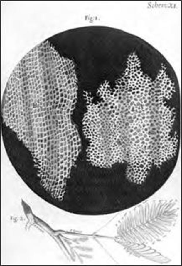
He searched for a name for them and finally decided on cells, from cella, a Latin word meaning "small room." (Hooke had not really seen "cells" but rather the outlines of walls that plant cells build around themselves; perhaps, nestled within them was an actual living cell, but there's no illustration that proves the point.) "A great many little boxes," as Hooke imagined them. Unwittingly, he had inaugurated a new conception of living beings, and of humans.
Hooke looked further and deeper for small, independent living units invisible to the naked eye. At a Royal Society assembly in November 1677, he described his microscopic observations on rainwater. The society recorded his observations:
The fi st experiment there exhibited was the pepper-water, which had been made with rain-water . . . put whole into it about nine or ten days before. In this Mr. Hooke had all week discovered great numbers of exceedingly small animals swimming to and fro. They appeared the bigness of a mite through a glass, that magnifi d about a hundred thousand times in bulk; and consequently, it was judged, that they were a hundred thousand times less than a mite. Their shape was to appearance like a very small clear bubble of an oval or egg form; and the biggest end of this egg-like bubble moved foremost. They were observed to have all manner of motions to and fro in the water; and by all, who saw them, they were verily believed to be animals; and that there could be no fallacy in the appearance.
In the decade that followed, Antonie van Leeuwenhoek, having learned of Hooke's earlier work, communicated with him, realizing that the animalcules that he had seen tumbling under his scopes might be analogous to the collection of living units—cells—that Hooke had witnessed in cork, or the organisms tumbling in pepper water. But there is an abject and disappointed tone in these letters, such as this one from November 1680: "As it has often reached my ear that I only tell fictitious stories about the little animals . . ." But in a prescient note, written in 1712, he continued, "Nay, we may yet carry it farther, and discover in the smallest particle of this little world a new inexhausted fund of matter, capable of being spun out into another universe."
Hooke replied only sporadically, but he ensured that Leeuwenhoek's letters were translated and presented to the Royal Society. Yet, although Hooke had likely saved Leeuwenhoek's reputation for posterity, his own influence on cell biological thinking was still rather limited. As the historian of cell biology Henry Harris described it: "Hooke did not for a moment suggest these structures were the residual skeletons of the basic subunits of which all plants and animals were constituted. Nor would he necessarily have imagined, if he had thought of basic subunits at all, that they would have the size and shape of the cork cavities that he had observed." He had seen "the walls of a living cell in cork, but he misunderstood their function, and he clearly had no conception of what, in the living state, occupied the spaces within these walls."* A piece of dead cork with pores in it; what more to make of his micrographic drawing? Why was a plant stem built in this manner? How did these "cells" arise? What was their function? Were they universal to all organisms? And what was the relevance of these living compartments to the normal body or to disease?
Hooke's interest in microscopy eventually dwindled. His peripatetic intellect needed to roam widely, and he returned to optics, mechanics, and physics. Indeed, Hooke's interest in virtually everything may have been his critical failing. The Royal Society's motto, Nullius in verba, translated loosely as "Take no one's word for evidence," was his personal mantra. He loped from one scientific discipline to the next, offering potent insights, believing no one's word, claiming dominion over critical parts of a science, but never asserting complete authority over any one subject. He had built himself on the model of the Aristotelian philosopher-scientist—an inquirer into all matters of the world, an adjudicator of all evidence—rather than the contemporary vision of the scientist as the authority on a single subject, and his reputation suffered as a result.
* In 1671, the Royal Society received two additional communications: one from the Italian scientist Marcello Malpighi and one from Nehemiah Grew, a secretary of the society, both describing cellular forms in various tissues, particularly in plant material. However, even though both Leeuwenhoek and Hooke acknowledged their work, both Malpighi's and Grew's observations on cellular anatomy were largely ignored in the seventeenth century. Grew's illustrations of cells in plant stems have been relegated to history, but Malpighi, who went on to explore the microscopic anatomy of animal tissues, lives on through many of the cellular structures that are named after him: among them, the Malpighian layer of the skin and the Malpighian cells in the kidney.
In 1687, Isaac Newton published Philosophiae Naturalis Principia Mathematica (Mathematical Principles of Natural Philosophy), a work so far-reaching in its depth and breadth that it shattered the past and shaped a new landscape for the future of science. Among its revelations: Newton's law of universal gravitation. Hooke, however, argued that he had formulated the laws of gravitation earlier, and that Newton had plagiarized his observations.
It was a preposterous claim. Indeed, Hooke, and several other physicists, had suggested that planetary bodies were attracted to the sun through invisible "forces," but none of the prior analysis had anywhere near the mathematical rigor or scientific depth that Newton brought to the puzzle in Principia. Hooke's and Newton's argument festered over decades, although Newton, arguably, had the last chortle. In one often-repeated story, likely apocryphal, the sole portrait of Hooke went missing when Newton oversaw the movement of the Royal Society to its new quarters in Crane Court in 1710, seven years after Robert Hooke's death—then neglected to commission a posthumous version. The pioneer of optics, the man who brought whole universes into view, is invisible to us. No definitive likeness, or portrait, of Hooke exists today.
From "The Song of the Cell: An Exploration of Medicine and the New Human" by Siddhartha Mukherjee. Copyright 2022 by Siddhartha Mukherjee, M.D. Reprinted by permission of Scribner, an imprint of Simon & Schuster, Inc.
For more info:
- "The Song of the Cell: An Exploration of Medicine and the New Human" by Siddhartha Mukherjee (Scribner), in Hardcover, eBook and Audio formats, available via Amazon, Barnes & Noble and Indiebound
- siddharthamukherjee.com
See also: